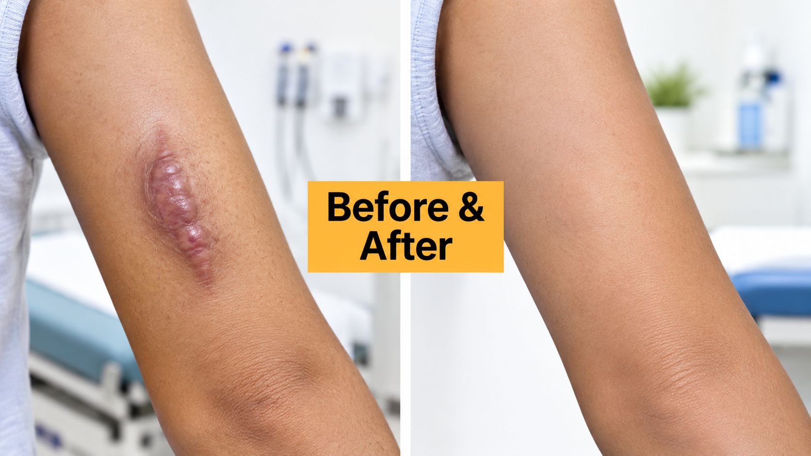
Keloid scars often look like a straightforward cosmetic problem. A raised, firm area is visible, the original injury may have healed long ago, and removal can seem like the obvious answer. Yet keloids behave differently from ordinary scars. They can extend beyond the original wound, remain itchy or tender, and return after treatment even when the early result looks smooth.
That makes keloid scar removal before and after more complicated than a single comparison photograph. The immediate “after” may show a flatter surface, while the meaningful clinical question is whether the scar stays controlled during healing. The examples below describe realistic treatment pathways and clinical scenarios used to explain decision-making at Chernoff Cosmetic Surgery. They aren't presented as undocumented individual patient records or guaranteed outcomes. Instead, they show how surgical, injection-based, laser, pressure, cryotherapy, and combination protocols can produce very different before-and-after journeys.
Table of Contents
- 1. Surgical Keloid Excision with Layered Closure Technique
- 2. Intralesional Corticosteroid Injections with Serial Treatments
- 3. Fractional CO2 Laser Resurfacing Combined with Surgical Excision
- 4. Silicone-Based Topical Treatments with Pressure Garments
- 5. Radiation Therapy Following Surgical Excision
- 6. Cryotherapy with Intralesional Steroid Combination
- 7. 5-Fluorouracil Intralesional Injections with Verapamil
- 7-Method Keloid Scar Removal: Before & After Comparison
- Choosing the Right Keloid Removal Path for You
1. Surgical Keloid Excision with Layered Closure Technique
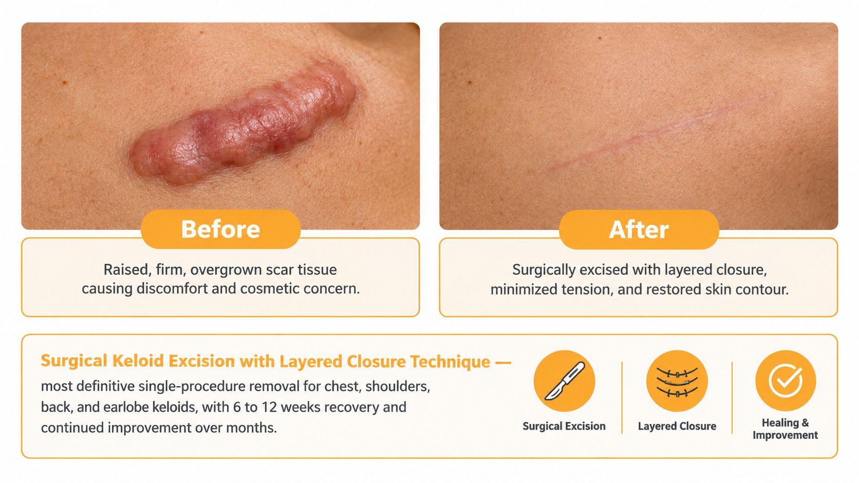
A large chest, shoulder, or upper-back keloid may need surgical removal when it is bulky, painful, repeatedly irritated, or difficult to manage with injections alone. The visible before-and-after change can be dramatic because the raised tissue is removed immediately. The important limitation is that excision alone isn't a complete keloid strategy.
Clinical reviews report recurrence after surgery alone in roughly 45% to 100% of cases, depending on the study and treatment context, as summarized by the American Academy of Family Physicians' clinical review of keloid management. A patient may therefore see a clean, flat early incision and still require a recurrence-prevention plan.
What the closure changes
Layered closure addresses one of the mechanical problems that can stimulate excessive scar formation. After removing the keloid tissue, the surgeon closes deeper tissue layers before bringing the skin edges together. Subcuticular sutures and carefully selected skin adhesives can help distribute tension rather than leaving the skin to bear all the pulling force.
This approach is especially relevant for a chest-wall keloid created after surgery or trauma, where movement and skin tension can challenge healing. An earlobe lesion may allow a more cosmetically refined closure, while a broad shoulder lesion may require a different reconstruction plan. The final scar won't look identical in every location.
Practical rule: A surgical before-and-after comparison should include the healing timeline, not just the first flat photograph.
Patients should plan for close follow-up and strict protection from friction, tension, and ultraviolet exposure. Depending on the case, a surgeon may add steroid injections, pressure therapy, radiation, or another adjunct rather than treating excision as a standalone procedure. Guidance on the broader principles of scar revision and treatment planning can help patients understand why closure technique is only one part of the result.
2. Intralesional Corticosteroid Injections with Serial Treatments
A smaller or actively growing keloid may be better suited to injections than immediate surgery. The clinician places a corticosteroid directly into the dense scar tissue, where it can reduce inflammatory activity and excessive collagen production. The result is gradual, not instantaneous. Over successive visits, the lesion may become softer, flatter, and less symptomatic.
This path is often practical for an earlobe keloid after piercing, an acne-related lesion on the chest, or a shoulder scar that remains raised but doesn't require bulk removal. It can also be useful after excision, when injections are used to manage early thickening at the incision edges. The treatment plan should be individualized because injection depth, concentration, spacing, and skin response all matter.
What patients should compare
A useful before-and-after record includes standardized photographs, symptom notes, and measurements of height and firmness. A scar that looks only modestly smaller may still be clinically improved if itching, tenderness, and tension have decreased. Conversely, a flatter lesion with visible lightening or indentation from treatment may need a change in technique.
Steroid injections aren't risk-free. Local skin thinning, pigment alteration, visible small blood vessels, and discomfort can occur, particularly when medication spreads beyond the intended scar. Patients with darker skin tones should discuss pigmentary changes before treatment, and the clinician should monitor the surrounding skin at each visit.
“The goal isn't simply to make the keloid smaller at the next appointment. The goal is to control its activity without creating a new cosmetic problem.”
Silicone products, pressure therapy, and careful sun protection may support the treatment plan, particularly when the scar is responding but remains vulnerable to irritation. Patients can review general ways to minimize scarring while following the specific instructions provided by their treating clinician.
3. Fractional CO2 Laser Resurfacing Combined with Surgical Excision
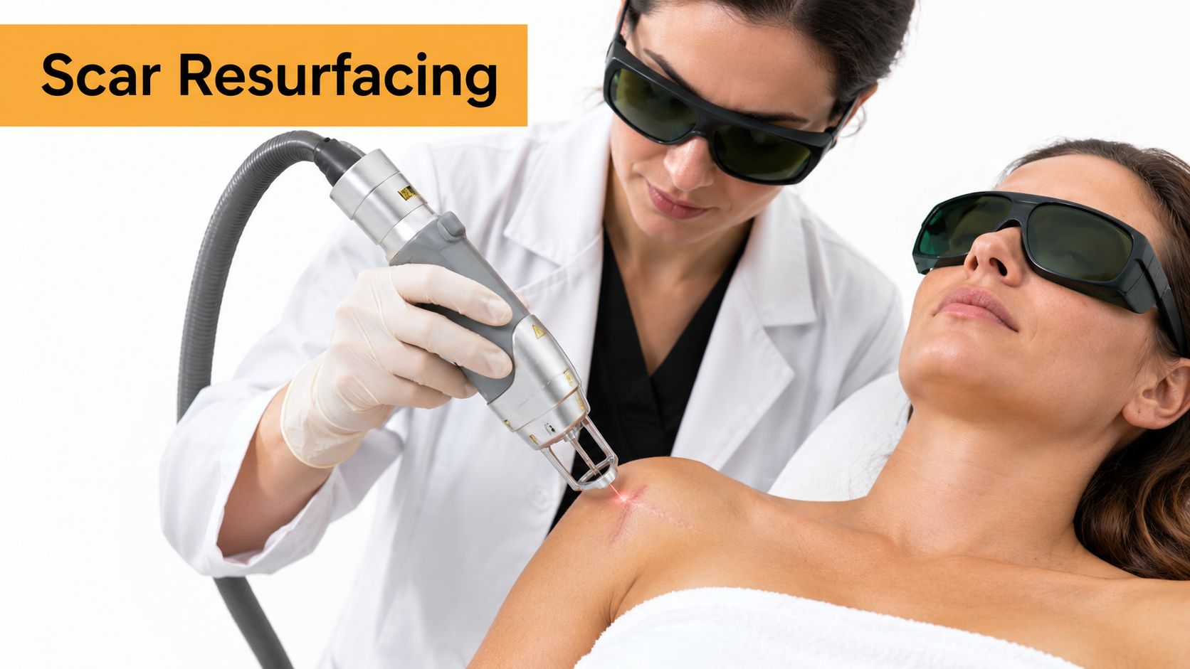
A shoulder keloid can be flatter after excision yet still leave a firm, irregular line. An earlobe lesion may have lost its raised tissue but retain uneven texture or color. At Chernoff Cosmetic Surgery, this before-and-after pathway pairs surgical removal with fractional CO2 laser resurfacing when the remaining concern is surface quality rather than scar bulk.
Surgical excision addresses the thicker tissue. Fractional CO2 laser treatment creates controlled microscopic treatment zones that can support remodeling in selected areas without removing the scar again. Patients can review this overview of fractional laser treatment for texture and surface refinement while discussing whether the timing and technique suit their scar.
The result is not guaranteed to be an invisible scar. Resurfacing may improve texture and blending, but it cannot remove an individual tendency to form keloids. The trade-off is that treatment must be selected carefully. A surface-focused procedure may not control an actively expanding keloid.
Timing and selection matter
Laser treatment is generally considered after the incision has healed sufficiently. The treating physician must determine whether the remaining mark is a stable postoperative scar, a recurrent keloid, or another raised scar. That distinction affects whether resurfacing is appropriate or whether the underlying activity needs attention first.
A useful before-and-after comparison includes the original lesion, the early surgical result, and later photographs taken under similar lighting. Height may improve before color does, while inflammation can make the scar look different during healing. Sun protection matters because recently treated skin may darken or become more noticeable after ultraviolet exposure.
A technique-specific series of surgical excision followed by superficial radiation reported remission in 39 of 48 patients, or 81%, while 9 patients, or 19%, were refractory, according to the published clinical case series. Those findings concern that combined radiation protocol, not fractional CO2 laser alone. Naming the adjunct prevents a before-and-after image from implying evidence for a treatment it did not evaluate.
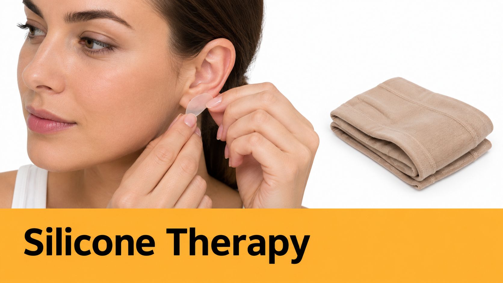
4. Silicone-Based Topical Treatments with Pressure Garments
Not every keloid needs an operation at the first sign of abnormal thickening. A small earlobe lesion, a newly thickened surgical scar, or a vulnerable incision may be managed conservatively with silicone and pressure when the clinician believes the approach is appropriate.
Silicone sheets and gels create an occlusive layer over fully closed skin. Pressure garments add sustained compression, which may reduce mechanical stimulation and help the scar mature more evenly. The combination is most useful when the patient can apply it consistently. Occasional use rarely provides a fair test of the protocol.
A realistic conservative pathway
Consider an early piercing-related earlobe keloid that has started to firm but hasn't become bulky. The patient may use a fitted pressure device with silicone after the skin is fully healed, while the clinician monitors thickness, color, itching, and extension beyond the original injury. The before-and-after result may be gradual flattening rather than complete disappearance.
Silicone isn't appropriate over an open, draining, or infected wound. Pressure also has to be comfortable and correctly fitted. Excessive compression can cause irritation, skin breakdown, or poor tolerance, while inadequate contact may provide little benefit.
- Apply consistently: Use the product according to the clinician's instructions and keep the skin clean and dry.
- Inspect the skin: Stop and seek advice if there is persistent redness, breakdown, rash, or increasing pain.
- Protect the area: Sun exposure can make a new scar more conspicuous, especially while the skin is still changing.
- Track the response: Photographs taken in consistent lighting are more useful than memory alone.
This path can also serve as maintenance after excision or injections. Its trade-off is time and adherence. A patient seeking an immediate reduction in a large, firm lesion may need a different approach, while someone with an early or prevention-focused concern may reasonably prefer a non-surgical plan.
5. Radiation Therapy Following Surgical Excision
Radiation is an adjunct, not a replacement for removing a bulky keloid. In selected cases, focused treatment is delivered soon after excision to reduce the activity of residual scar-forming cells. The timing matters because the wound is entering the phase in which recurrent keloid growth can begin.
This pathway may be discussed for a recurrent chest-wall keloid, a difficult shoulder lesion, or an earlobe keloid that returned after an earlier excision. It requires coordination between the surgeon and radiation specialists, along with a clear discussion of risks, benefits, wound care, and long-term follow-up.
Why the protocol changes the comparison
The most misleading before-and-after image is a photograph of a flat incision labeled as a successful removal without identifying the postoperative treatment. A systematic review of surgical management for major keloids describes recurrence around 21% across 244 cases and reports that outcomes varied according to wound coverage and postoperative radiation. In one subgroup, wound coverage with postoperative radiation had 0% recurrence, compared with 16% for excision plus radiation alone. These findings apply to the reviewed treatment groups and shouldn't be promised to an individual patient.
Radiation decisions must account for lesion location, prior treatment, age, medical history, and the amount of tissue involved. A consultation should explain why radiation is being considered, what schedule is proposed, and which side effects deserve attention. It also shouldn't be presented as a universal answer for every keloid.
The durable “after” is often the result of coordinated timing, not the excision itself.
Pressure therapy, silicone, injections, and follow-up may still form part of the plan. Patients should ask who will coordinate the treatment and how recurrence will be assessed after the wound appears healed.
6. Cryotherapy with Intralesional Steroid Combination
Cryotherapy can be useful for selected raised scars, particularly when reducing bulk without making a conventional incision is a priority. Controlled freezing damages scar tissue and can soften the lesion. A corticosteroid injection may then help suppress continued collagen production and inflammatory activity.
An earlobe keloid after piercing is a familiar clinical scenario. A patient may have a firm, rounded lesion that is too established for topical care alone but small enough for serial office treatment. Another scenario involves multiple small acne-related keloids on the chest or back, where treating each lesion individually may be more practical than performing broad excision.
The before-and-after is gradual
After treatment, the area may become swollen, blistered, crusted, or temporarily darker or lighter. The immediate appearance shouldn't be mistaken for the final result. The clinician needs to assess how the tissue heals before deciding whether another session is appropriate.
Cryotherapy carries a particular pigmentary trade-off. Melanin-producing cells can be sensitive to cold injury, so hypopigmentation may be a significant concern for some patients. A careful discussion of skin type, lesion location, freeze technique, wound care, and the possibility of repeat treatment is essential.
Patients should keep the area clean, avoid picking at blisters, and follow the prescribed dressing instructions. Sun protection becomes important once the surface has healed. Pressure therapy or silicone may be added between sessions when appropriate, especially if the lesion remains vulnerable to friction or tension.
The best comparison photographs show the original height and width, the healing phase, and the later contour. They should also record symptoms. A lesion that is smaller but still itchy may need continued management, while a scar with less pain and a softer texture may represent meaningful progress even before the color normalizes.
7. 5-Fluorouracil Intralesional Injections with Verapamil
Some keloids remain active despite conventional injections or return after surgery. In that setting, a clinician may discuss less routine intralesional medication protocols, including combinations such as 5-fluorouracil and verapamil. These medications act through different pathways related to fibroblast behavior and collagen production, but their use requires careful patient selection and experienced administration.
A representative scenario is a recurrent chest keloid after a previous excision and steroid treatment. Another involves a thick earlobe lesion that has not responded adequately to standard injection care. The aim may be reduction in firmness, thickness, and symptoms rather than an instant cosmetic transformation.
Advanced treatment needs careful monitoring
The clinician should document the lesion before treatment, including its dimensions, depth, color, symptoms, and prior therapies. Patients need to understand that this isn't automatically a first-line option and that local reactions, discomfort, ulceration, pigment changes, or other complications can occur.
A sound consultation covers medication history, allergies, pregnancy considerations where relevant, immune status, and previous responses. Treatment intervals and dosage should be determined by the treating clinician rather than copied from another patient's plan. Cold compresses, pressure therapy, and specific wound-care instructions may be recommended after injection.
Clinical perspective: A recurrent keloid is a treatment history, not just a raised mark. Previous surgery, injections, radiation, pressure, and healing behavior all influence the next decision.
Before-and-after photographs should be paired with objective clinical notes. A softer lesion may be improving even if its outline remains visible, while a rapidly enlarging area needs prompt reassessment. Patients should also ask whether the proposed protocol is intended to control recurrence, reduce symptoms, improve contour, or address all three goals.
7-Method Keloid Scar Removal: Before & After Comparison
| Treatment | Implementation complexity | Resource requirements | Expected outcomes | Ideal use cases | Key advantages |
|---|---|---|---|---|---|
| Surgical Keloid Excision with Layered Closure Technique | High, surgical skill and anesthesia required | Operating room or procedure suite, experienced surgeon, sutures, adhesives, postop care | Immediate keloid removal with visible improvement in 6–12 weeks; recurrence risk if no adjunctive therapy | Large keloids (chest, shoulders, back), contour revision, earlobe keloids | Most definitive single-procedure removal; allows scar revision and contouring |
| Intralesional Corticosteroid Injections with Serial Treatments | Low–Moderate, office procedure with repeated visits | Clinic setting, triamcinolone, trained injector, serial follow-up | Gradual flattening/softening over 3–6 months; variable response (≈50–80%); possible recurrence | Small–moderate keloids, difficult locations, first-line or adjunctive therapy | Non-invasive, low downtime, cost-effective, adjustable treatment intensity |
| Fractional CO2 Laser Resurfacing Combined with Surgical Excision | High, multimodal technique requiring laser and surgical expertise | OR/clinic, fractional CO2 laser, trained operator, staged sessions, postop protocol | Improved texture and color with continued remodeling over 6–12 months; superior cosmetic results | Patients needing excision plus maximal cosmetic refinement or pigment/texture issues | Combines removal with collagen remodeling for enhanced aesthetic outcomes |
| Silicone-Based Topical Treatments with Pressure Garments | Low, home-based but requires consistent adherence | Pharmaceutical-grade silicone products, fitted compression garments, patient compliance | Gradual improvement over 3–6 months; best for prevention and maintenance; modest on established large keloids | Early-stage keloids, post-op prevention, long-term maintenance | Lowest cost, no downtime, safe for all skin types, easy at-home use |
| Radiation Therapy (Brachytherapy) Following Surgical Excision | High, requires coordinated multidisciplinary care | Radiation oncology equipment, timing within 24–48 hours post-op, specialist team | Markedly reduced recurrence (<15% vs ~45% without adjunct); outcomes assessed at 6–12 months | High-risk or recurrent keloids (chest, shoulders, ears), extensive disease after excision | Most effective prophylactic adjunct to prevent recurrence after surgery |
| Cryotherapy (Liquid Nitrogen) with Intralesional Steroid Combination | Low–Moderate, office-based but needs technique to avoid complications | Cryotherapy device/liquid nitrogen, steroids, multiple short sessions | Rapid flattening over weeks to months; blistering and pigment changes possible; multiple sessions required | Earlobe and small-to-moderate keloids, patients avoiding surgery | Quick, affordable, dual mechanism increases efficacy versus single modality |
| 5-Fluorouracil (5-FU) Intralesional Injections with Verapamil | Moderate–High, specialized pharmacologic administration and monitoring | 5-FU and verapamil supplies, experienced clinician, careful dosing and follow-up | Effective reduction in resistant or recurrent keloids over 2–4 months; multiple injections needed | Treatment-resistant or recurrent keloids, large or difficult-to-access lesions | Targets cellular pathways of keloid formation; effective when other therapies fail |
Choosing the Right Keloid Removal Path for You
The right treatment depends on more than the size of the scar. Location, skin type, symptoms, wound tension, previous recurrence, and the patient's ability to follow a long aftercare plan all influence the recommendation.
A small, early keloid on the earlobe may be approached with injections, silicone, pressure, cryotherapy, or a combination. A broad, firm chest or shoulder lesion may need excision followed by an adjunct designed to reduce recurrence. A recurrent lesion deserves a different conversation from a first-time lesion because the previous response provides important information about its biology.
The location also changes the cosmetic and mechanical trade-offs. Earlobe tissue may allow refined closure and pressure therapy. The chest, shoulder, and upper back experience more movement and tension, which can make recurrence prevention particularly important. Scars near the jawline or other visible areas may require careful discussion of color, texture, and the patient's tolerance for staged treatment.
The American Academy of Dermatology notes that keloids can return after surgical removal and that treatment often combines approaches such as injections, pressure, silicone, or radiation. Its patient guidance on keloid treatment reinforces a point that every before-and-after gallery should make clear: an early improvement isn't the same as permanent control.
At consultation, ask:
- What is the diagnosis: Is the lesion a keloid, a hypertrophic scar, or another raised growth?
- What is the recurrence plan: If excision is recommended, which adjunct will follow it and why?
- What will healing look like: How should you respond to swelling, blistering, pigment change, drainage, or renewed growth?
- How will progress be measured: Will the team use photographs, dimensions, symptom tracking, or all three?
- What happens if it returns: Is there a planned next step rather than an assumption that the first treatment will be definitive?
A recent meta-analysis of major keloid treatment reported an average ear-keloid recurrence of 20.3% across 26 studies and discussed differences related to location and treatment selection in the PubMed review. That figure describes pooled research, not an individual prediction. It does show why the best treatment plan is location-specific rather than built around a single universal procedure.
Chernoff Cosmetic Surgery can evaluate the scar's history and coordinate surgical, injection, laser, and adjunctive options when appropriate. The goal is a realistic before-and-after plan that accounts for both the visible contour and the months of follow-up needed to protect the result.
Chernoff Cosmetic Surgery offers individualized evaluation for keloid scars, including surgical removal and non-surgical scar treatments selected around the lesion's location, history, and recurrence risk. Visit Chernoff Cosmetic Surgery to request a consultation and discuss which treatment path best fits your before-and-after goals.